43 diagram of the lungs with labels
Occipital lobe: Anatomy, function and clinical relations - Kenhub The occipital lobe is the visual processing area of the brain. Brodmann areas 17,18 and 19 are located within the occipital lobe and form the visual cortices. Brodmann area 17 (V1) forms the primary visual cortex while V2,V3,V4, or Brodmann areas 18 and 19 form the visual association cortex. What is the Circumflex Artery? (with pictures) - Info Bloom The circumflex artery is a branch of the left coronary artery in the heart. Also known as the LCX, it is situated to the left atrium on the outside of the heart wall. This blood vessel branches off the left coronary artery on the top left side of the heart. The coronary artery arises from the aorta, the major artery exiting the heart to deliver ...
Pulmonary edema - Symptoms and causes - Mayo Clinic Blood clot in the lungs (pulmonary embolism). A blood clot moving from the blood vessels in the legs to the lungs can cause pulmonary edema. Exposure to certain toxins. Inhaling toxins or breathing in some stomach contents when vomiting (aspiration) causes intense irritation of the small airways and air sacs, resulting in fluid buildup.
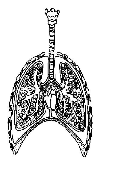
Diagram of the lungs with labels
Biology Diagrams Images Pictures Of Human Anatomy And Physiology --> Biology Clipart Human Physiology Biology Human Physiology Transparent Browse 5,145 professional human anatomy physiology stock photos available royalty free Path of Blood Through the Heart - New Health Advisor Blood circulates in the lungs, where oxygen (O2) is added to the bloodand carbon dioxide (CO2) is removed. Blood returns to the heart through the pulmonary veins, which go into the left chambers. 4. Left Atrium. The left atrium receive blood from the lungs and pushes it through the mitral valve to pass it into the left ventricle. 5. Left Ventricle How to Use Your Peak Flow Meter - Asthma Initiative of Michigan Take a deep breath. Fill your lungs all the way. Hold your breath while you place the mouthpiece in your mouth, between your teeth. Close your lips around it. Do not put your tongue inside the hole. Blow out as hard and fast as you can. Your peak flow meter will measure how fast you can blow out air. Write down the number you get.
Diagram of the lungs with labels. Digestive System - Lesson - TeachEngineering The digestive system is amazing: it takes the foods we eat and breaks them into smaller components that our bodies can use for energy, cell repair and growth. This lesson introduces students to the main parts of the digestive system and how they interact. In addition, the associated activity helps students learn about some of the challenges astronauts face when eating in outer space. Engineers ... reference.yourdictionary.com › resources › parts-ofParts of the Body for Kids: Names & Basic Functions protecting lungs and heart; assisting in arm movement. abdomen. middle of the torso. stabilizing the body; protecting internal organs. belly button. middle of the belly. connects a baby’s umbilical cord to its mother’s placenta before birth. back. back of the torso. stabilizing the body; protecting vertebrae, which send messages to the rest ... Immune system - Wikipedia The immune system is a network of biological processes that protects an organism from diseases.It detects and responds to a wide variety of pathogens, from viruses to parasitic worms, as well as cancer cells and objects such as wood splinters, distinguishing them from the organism's own healthy tissue.Many species have two major subsystems of the immune system. Blood Cell Basics - Activity - TeachEngineering Blood is made up of many different things: plasma (5%), water (50%), white blood cells and platelets (1%), and red blood cells (44%). The plasma is a water and salt substance that contains the blood's proteins and suspends the cells. The white blood cells are known as the army and fight off infections and diseases.
Circulatory system - Wikipedia The circulatory system includes the heart, blood vessels, and blood. The cardiovascular system in all vertebrates, consists of the heart and blood vessels. The circulatory system is further divided into two major circuits - a pulmonary circulation, and a systemic circulation. The pulmonary circulation is a circuit loop from the right heart taking deoxygenated blood to the lungs where it is ... Pleural Cavity: Anatomy, Effusion Causes, Treatment - Verywell Health The pleural cavity is the space that lies between the pleura, the two thin membranes that line and surround the lungs.The pleural cavity contains a small amount of liquid known as pleural fluid, which provides lubrication as the lungs expand and contract during respiration.When increased fluid occupies this space, it is referred to as a pleural effusion and can severely restrict breathing ... Hands-on Activity Polluted Air = Polluted Lungs - TeachEngineering.org Worksheet: To review the primary respiratory system components, have students label parts and describe their functions on the Respiratory System Worksheet. After students are finished, have them volunteer to write the parts and functions on a larger diagram to post in the classroom, or on a projected overhead transparency of the worksheet. en.wikipedia.org › wiki › AcetylcholinesteraseAcetylcholinesterase - Wikipedia Acetylcholinesterase (HGNC symbol ACHE; EC 3.1.1.7), also known as AChE, AChase or acetylhydrolase, is the primary cholinesterase in the body. It is an enzyme that catalyzes the breakdown of acetylcholine and some other choline esters that function as neurotransmitters.
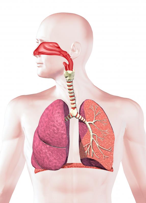
Trunk Region (Torso) < Regional Anatomy - Wellness Advocate . com Trunk (Torso) In our body, the Trunk Region (Torso), an anatomical term for the central part of the body, is a combination of both the thoracic region (chest), including the mammary region (breasts), and the abdomen region (belly region) including the naval (umbilicus region), coxal region, and pubic region. In our body, the Trunk Region (Torso) as an anatomical term for the central part of ... Positions and Functions of the Four Brain Lobes - MD-Health.com The occipital lobe, the smallest of the four lobes of the brain, is located near the posterior region of the cerebral cortex, near the back of the skull. The occipital lobe is the primary visual processing center of the brain. Here are some other functions of the occipital lobe: Visual-spatial processing. Movement and color recognition. Respiratory System Organs and Their Functions - New Health Advisor The three major parts of the respiratory system all work together to carry out their task. The airways (nose, mouth, pharynx, larynx etc.) allow air to enter the body and into the lungs. The lungs work to pass oxygen into the body, whilst removing carbon dioxide from the body. The muscles of respiration, such as the diaphragm, work in unison to ... Respiratory System (Theory) : Rodent Anatomy - Amrita Vishwa Vidyapeetham A pair of lungs is enclosed in the thoracic cavity. The ventilating system consists of the thoracic cage and intercostals muscles, diaphragm, and elastic connecting tissue of the lung which move air within the respiratory tract. The air from outside passes through the external nostrils and nasal passages into pharynx.
Respiratory System Worksheet - WikiEducator
Creating Model Working Lungs: Just Breathe - TeachEngineering.org A model of the lungs. Pull the diaphragm (balloon) down (that is, away from the lungs) in order to inflate the lungs. (Note: This makes the chest cavity larger and decreases the pressure.) Push the diaphragm (balloon) in (towards the lungs) in order to deflate the lungs.
Respiratory system jnr
Circulatory System Diagram | New Health Advisor It covers the movement of blood from heart to the lungs where the oxygenation of blood takes place. In oxygenation the waste material and carbon dioxide are removed from the blood while oxygen is taken up by the red blood cells. ... There are different types of circulatory system diagrams; some have labels while others don't. The color blue ...
What is Rheumatoid Arthritis of the Lung? (with pictures)
Diagram of Human Heart and Blood Circulation in It - New Health Advisor Four Chambers of the Heart and Blood Circulation. The shape of the human heart is like an upside-down pear, weighing between 7-15 ounces, and is little larger than the size of the fist. It is located between the lungs, in the middle of the chest, behind and slightly to the left of the breast bone. The heart, one of the most significant organs ...

muscular system worksheets - Bing Images | School for me ... | Human respiratory system ...
Brain, spinal cord and peripheral nervous system anatomy - Kenhub Diagram of the brain (labeled) The most superficial layer of the cerebrum is the cerebral cortex.It is a layer of grey matter which displays numerous folds (sulci and gyri), can be categorization structurally (cortical cytoarchitecture) or functionally (Brodmann areas), and is home to areas such as the primary motor cortex and the primary somatosensory cortex, both of which house a homunculus.
Respiration Worksheet Answers - WikiEducator
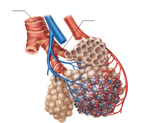
Human respiratory system stock photos and images (9,650) Human full respiratory system cross section Stock Images by Pixelchaos 90 / 3,350 The respiratory system, labeled Stock Photography by alila 54 / 2,728 Pulmonar alveoli Pictures by Andreus 16 / 300 Human Respiratory System Picture by nerthuz 9 / 245 Human respiratory system Stock Images by bluering 2 / 317 Human respiratory system Picture by ...

Respiratory System
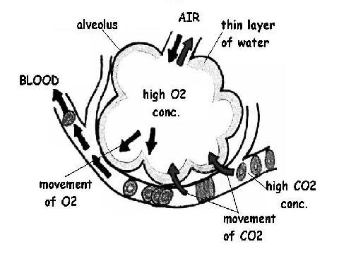
Mechanism of Respiration in Human Beings - Embibe Exams The mechanics of respiration in human beings takes place in two events: inspiration and expiration (inhale and exhale). The respiratory mechanism is the act of inhaling air into the lungs and is known as inspiration. Expiration is the process of removing air from the lungs. Amoeba and other unicellular organisms breathe through their skin.
Post a Comment for "43 diagram of the lungs with labels"