43 microscope images with labels
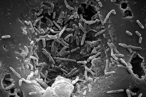
50 Striking Microscopic Images of Viruses and Bacteria March 07, 2022. 1/50. Colorized transmission electron micrograph showing H1N1 influenza virus particles. Surface proteins on the virus particles are shown in black. (Credit: NIAID) Bacteria and ... Cecum Histology Slide with Labeled Image and Diagram The tunica muscular layer of the provided cecum labeled image shows two distinct smooth muscle layers - inner longitudinal or oblique bundles and outer wavy bundles. Again, the cecum images show some elastic fibers in this layer. In addition, the cecum labeled image shows a thin and loose connective tissue layer with numerous blood vessels.
Light Microscope- Definition, Principle, Types, Parts, Labeled Diagram ... A light microscope is a biology laboratory instrument or tool, that uses visible light to detect and magnify very small objects and enlarge them. They use lenses to focus light on the specimen, magnifying it thus producing an image. The specimen is normally placed close to the microscopic lens.

Microscope images with labels
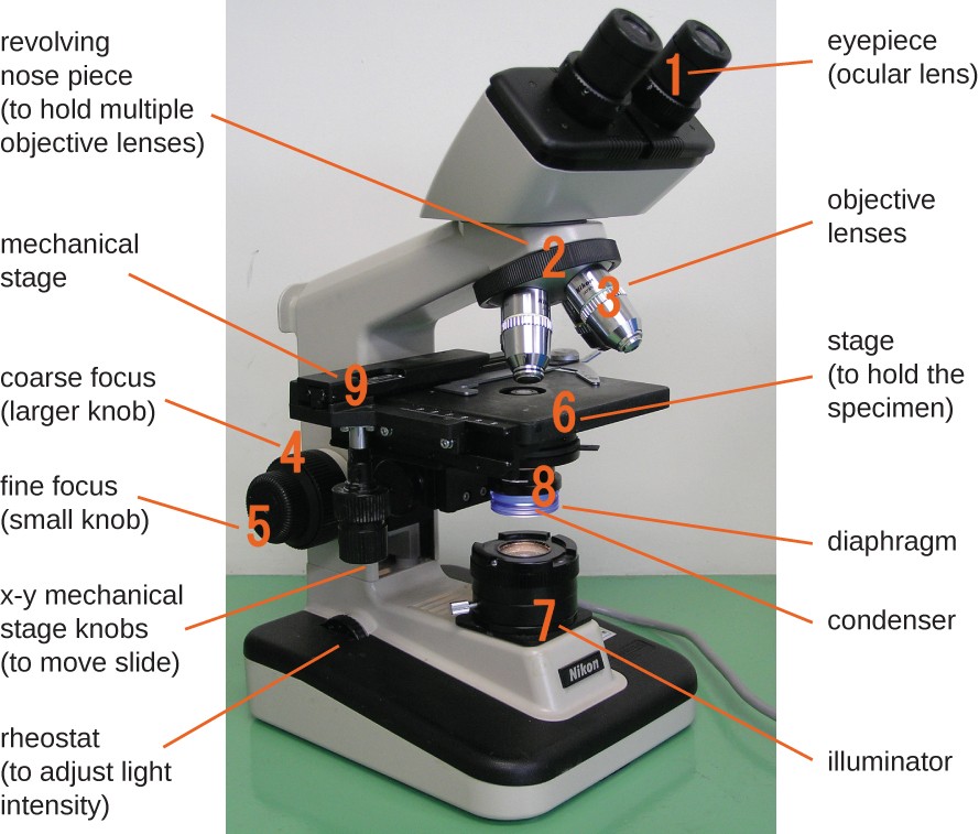
Bright-field microscope (Compound light microscope) - Diagram (Parts ... Bright-field microscope parts (Labeled Diagram) Ocular Lens This microscope has two eye lenses or ocular lens on the top of the microscope that are used to focus the image from the objective lens. It is from these lenses that we see the magnified image of the specimen. Objective Lens Binocular Microscope Anatomy - Parts and Functions with a Labeled ... Now, I will discuss the details anatomy of the light compound microscope with the labeled diagram. Why it is called binocular: because it has two ocular lenses or an eyepiece on the head that attaches to the objective lens, this ocular lens magnifies the image produced by the objective lens. Binocular microscope parts and functions Compound Microscope - Diagram (Parts labelled), Principle and Uses Compound Microscope - Diagram (Parts labelled), Principle and Uses As the name suggests, a compound microscope uses a combination of lenses coupled with an artificial light source to magnify an object at various zoom levels to study the object. A compound microscope: Is used to view samples that are not visible to the naked eye
Microscope images with labels. Electron Microscope- Definition, Principle, Types, Uses, Labeled Diagram There are two types of electron microscopes, with different operating styles: 1. Transmission Electron Microscope (TEM) The transmission electron microscope is used to view thin specimens through which electrons can pass generating a projection image. The TEM is analogous in many ways to the conventional (compound) light microscope. Researchers demonstrate label-free super-resolution microscopy Optics & Photonics April 21, 2022 Researchers demonstrate label-free super-resolution microscopy by Optica Researchers have developed a new measurement and imaging approach that can resolve... Microscope Parts | A Guide on their Location and Function The image of a compound microscope with labeled parts. Eyepiece. It is the part that you encounter when viewing an object in the microscope from the top. This is the first lens that helps to magnify the image. Based on the magnification power, the lens can be of 5X, 10X, 15X, or more. Parts of the Microscope with Labeling (also Free Printouts) Microscopes are specially created to magnify the image of the subject being studied. This exercise is created to be used in homes and schools. the microscope layout, including the blank and answered versions are available as pdf downloads. Click to Download : Label the Parts of the Microscope (A4) PDF print version.
Microscope Parts, Function, & Labeled Diagram - slidingmotion Microscope parts labeled diagram gives us all the information about its parts and their position in the microscope. Microscope Parts Labeled Diagram The principle of the Microscope gives you an exact reason to use it. It works on the 3 principles. Magnification Resolving Power Numerical Aperture. Parts of Microscope Head Base Arm Eyepiece Lens Electron Microscope Principle, Uses, Types and Images (Labeled Diagram ... Electron Microscope Principle, Uses, Types and Images (Labeled Diagram), Price Electron Microscope The advances in technology have enabled the development of powerful microscopes to view the samples at a nanometer level and thus were born the electron microscopes. The Best Photos Taken Through Microscopes Will Blow You Away Since 1974, Nikon has held a photography competition to recognize excellent images taken with the assistance of a microscope. In 2021, the competition received almost 2,000 entries from 88 countries. In these images, art and science come together in a surprising and beautiful way. We looked through this year's winning images to share our favorites. Brightfield Microscope (Compound Light Microscope)- Definition ... Brightfield Microscope is also known as the Compound Light Microscope. It is an optical microscope that uses light rays to produce a dark image against a bright background. It is the standard microscope that is used in Biology, Cellular Biology, and Microbiological Laboratory studies. This microscope is used to view fixed and live specimens ...
Parts of a microscope with functions and labeled diagram Optical parts of a microscope and their functions The optical parts of the microscope are used to view, magnify, and produce an image from a specimen placed on a slide. These parts include: Eyepiece - also known as the ocular. This is the part used to look through the microscope. Its found at the top of the microscope. Blood Histology Slides with Description and Labeled Diagram The blood is a specialized connective tissue that is fluid and circulates through the vascular channel. In the blood histology slide, you will find different types of cells with their specific features. This might be a short article where I will show you all the cells from the blood microscope slide with a labeled diagram and actual pictures. Microscope- Definition, Parts, Functions, Types, Diagram, Uses It is a type of fluorescence microscope that is used to produce 2-D or 3-D images of relatively thick specimens. In this type, the excitation light is focused on a specific spot of sample lying on the focal plane. The focus spot is optically manipulated to scan the entire sample and generate a 3-D image. Microscopic Morphology - BIO 2410: Microbiology - Baker College What are the shapes of the bacteria labeled 1 and 3 in this image? Shape 5A A1 Bacillus (streptobacillus) A3 Bacillus . Shape 6 1. What is the shape of the bacteria labeled in this image? ... The microscope images in this section show different bacterial structures visible using the light microscope. All images were photographed at 1000x ...
32 Label Of Compound Microscope - Label Design Ideas 2020
Microscope Types (with labeled diagrams) and Functions This is an advanced microscope that has specific application in viewing, observing and measuring the optical thickness and phase of completely transparent specimens and objects. A tiny interferometer is used and a specimen is placed on beam path of it. This path is split and then rejoined to create two superimposed images of the specimen in focus.
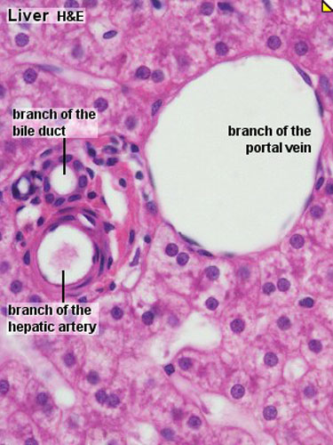
ANAT2241 Liver, Gallbladder, and Pancreas - Embryology
Mushroom Microscope Images, Stock Photos & Vectors | Shutterstock 1,335 mushroom microscope stock photos, vectors, and illustrations are available royalty-free. See mushroom microscope stock video clips. of 14. paint under microscope microscope food electron microscope images mold microscope mycelium bacteria microscope. Try these curated collections. Search for "mushroom microscope" in these categories. Next ...
23 Label And Color The Parts Of Both Microscopes - Labels 2021
Simple Microscope - Diagram (Parts labelled), Principle, Formula and Uses A simple microscope consists of Optical parts Mechanical parts Labeled Diagram of simple microscope parts Optical parts The optical parts of a simple microscope include Lens Mirror Eyepiece Lens A simple microscope uses biconvex lens to magnify the image of a specimen under focus.

31 Label The Indicated Parts Of The Microscope - Labels For Your Ideas
Compound Microscope - Types, Parts, Diagram, Functions and Uses Eyepiece/ocular lens - It is the part of the microscope that is looked through at the top. It comes with a magnification ranging between 5x and 30x. Image 3: The head connects the eyepiece to the objective lens. Picture Source: microscope.com. Head (monocular/binocular) - It is the structural support of the microscope.

MICROBIOLOGY SLIDE SPECIMENS
Microscope, Microscope Parts, Labeled Diagram, and Functions Microscope, Microscope Parts, Labeled Diagram, and Functions What is Microscope? A microscope is a laboratory instrument used to examine objects that are too small to be seen by the naked eye. It is derived from Ancient Greek words and composed of mikrós, "small" and skopeîn,"to look" or "see".

Microscope labeling
Sperm Under Microscope with Labeled Diagram Sperm Under Microscope 400X Labeled Diagram Before that, you may also read the below-mentioned article to get a full idea of the structure of seminiferous tubules - Histological features of the seminiferous tubules with the labeled diagram Okay, first, let's see the different histological features of the seminiferous tubules of an animal.

Microscope labeling
Simple Microscope - Parts, Functions, Diagram and Labelling Simple Microscope - Parts, Functions, Diagram and Labelling A microscope is one of the commonly used equipment in a laboratory setting. A microscope is an optical instrument used to magnify an image of a tiny object; objects that are not visible to the human eyes. Table of Contents The common types of microscopes are: What is a Simple microscope?
Mycobacterium leprae* - microbewiki
Compound Microscope- Definition, Labeled Diagram, Principle, Parts, Uses In order to ascertain the total magnification when viewing an image with a compound light microscope, take the power of the objective lens which is at 4x, 10x or 40x and multiply it by the power of the eyepiece which is typically 10x. Therefore, a 10x eyepiece used with a 40X objective lens will produce a magnification of 400X.

Best Top Desktop Wallpapers: Electron microscope images
Simple Squamous Epithelium under a Microscope with a Labeled Diagram Histological features of lung parenchyma with microscopic slide images and labeled diagrams. The lung's alveoli give the honeycomb appearance in the parenchyma and lines by flattened simple squamous epithelium. These alveoli are thin-walled and fills with air. From the lung parenchyma labeled diagram, you might identify the following structures -

Labels Of The Microscope
Electron Microscopy Images - Dartmouth We have a library of images recorded using our scanning and transmission electron microscopes. Some are shown below and others elsewhere. These images are in the public domain. If you have questions about the images or want some specific images contact Max Guinel . Hibiscus Flower (August 2021) Morphy Amorphophallus titanum anther cross section.
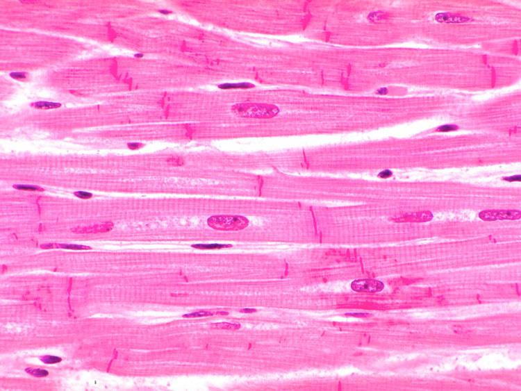
Tissues Flashcards | Easy Notecards
Compound Microscope - Diagram (Parts labelled), Principle and Uses Compound Microscope - Diagram (Parts labelled), Principle and Uses As the name suggests, a compound microscope uses a combination of lenses coupled with an artificial light source to magnify an object at various zoom levels to study the object. A compound microscope: Is used to view samples that are not visible to the naked eye

Microscope Clip Art at Clker.com - vector clip art online, royalty free & public domain
Binocular Microscope Anatomy - Parts and Functions with a Labeled ... Now, I will discuss the details anatomy of the light compound microscope with the labeled diagram. Why it is called binocular: because it has two ocular lenses or an eyepiece on the head that attaches to the objective lens, this ocular lens magnifies the image produced by the objective lens. Binocular microscope parts and functions
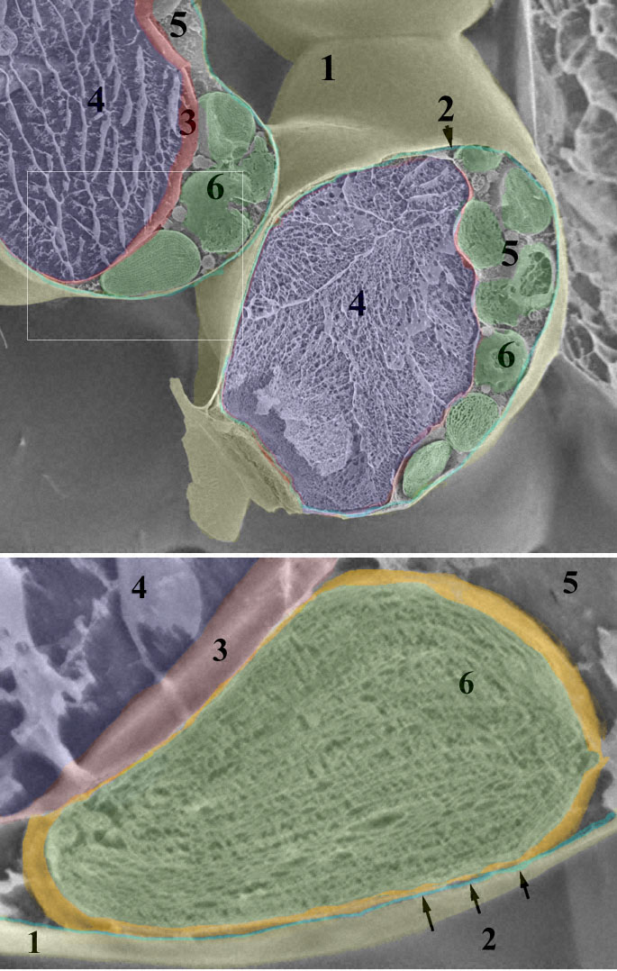
Leaf chloroplast
Bright-field microscope (Compound light microscope) - Diagram (Parts ... Bright-field microscope parts (Labeled Diagram) Ocular Lens This microscope has two eye lenses or ocular lens on the top of the microscope that are used to focus the image from the objective lens. It is from these lenses that we see the magnified image of the specimen. Objective Lens
Microscope Introduction – “e” Lab - Biology LibreTexts

Tongue Histology | Histology slides, Human anatomy and physiology, Tissue biology
32 Picture Of Microscope With Label - Labels For You

Labeling a Compound Microscope Quiz
Post a Comment for "43 microscope images with labels"