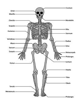
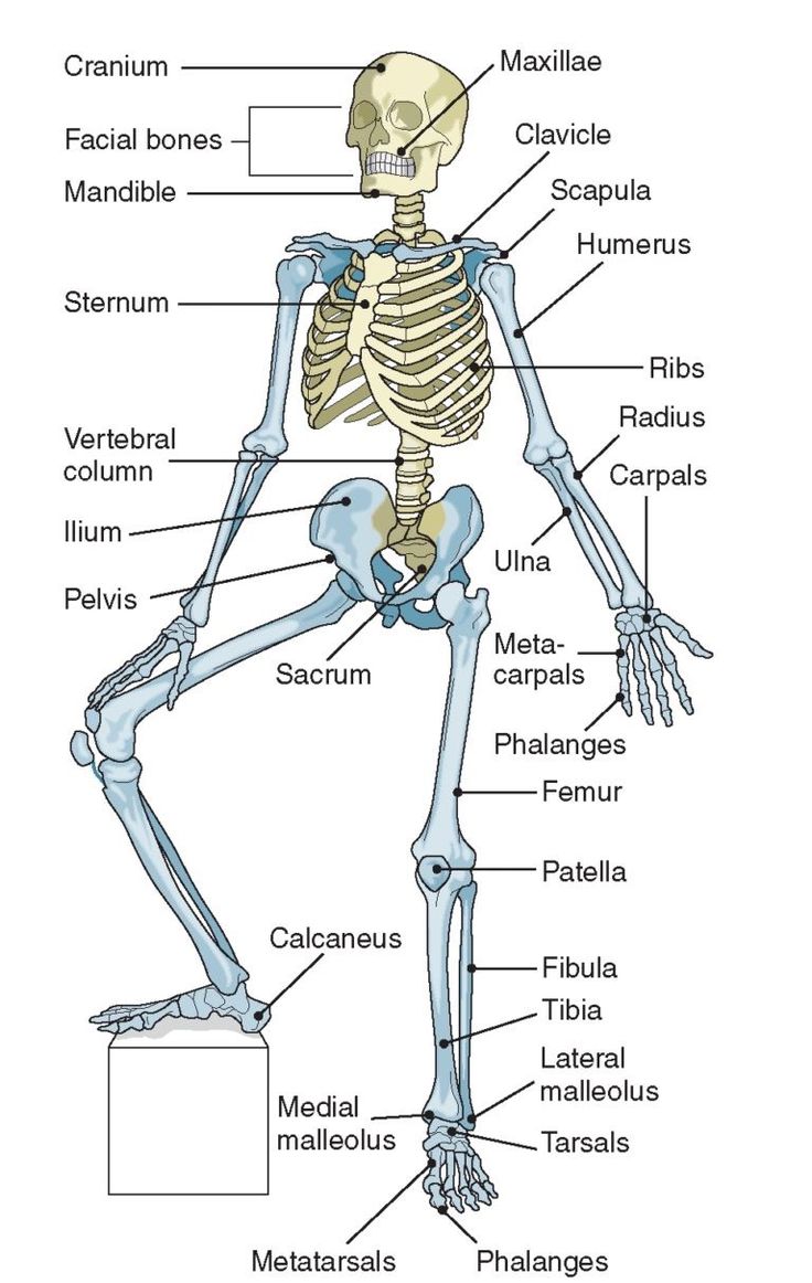
44 skeletal system diagram with labels
≡ 20 Important Organs of the Human Body List + Size & Weight 3.1. Upper right abdominal cavity just beneath the diaphragm. The liver ("Iecur" or "hepar") is the largest gland in the body with soft smooth surface, left and right lobes and weighs between 1.4-2 kg (3-4.4 lbs). The tissue consists of around 100,000 lobules and is metabolically the most complex human internal organ. Mechanisms of bone pain: Progress in research from bench to bedside Anatomically, sensory pathway in vertebrates consists of peripheral nervous system (PNS) and central nervous system (CNS). Primary afferent neurons in the dorsal root ganglion (DRG) and trigeminal...
Positions and Functions of the Four Brain Lobes - MD-Health.com The occipital lobe, the smallest of the four lobes of the brain, is located near the posterior region of the cerebral cortex, near the back of the skull. The occipital lobe is the primary visual processing center of the brain. Here are some other functions of the occipital lobe: Visual-spatial processing. Movement and color recognition.
Skeletal system diagram with labels
Cardiovascular System Worksheet Pdf - flakeinspire Cardiovascular system diagrams, quizzes and free worksheets. 1 cardiovascular system components of the cardiovascular system • consists of the heart plus all the blood vessels • transports blood to all parts of the body in two 'circulations': P 14 of a study guide for 4th grade via westerndairyassociation org wp wp 06 skeletal system pdf ... Sequence Diagram Tutorial - Complete Guide with Examples A sequence fragment is represented as a box that frames a section of interactions between objects (as shown in the examples below) in a sequence diagram. It is used to show complex interactions such as alternative flows and loops in a more structured way. On the top left corner of the fragment sits an operator. Functions of Human Skeletal System | Just-Health.net It is part of the skeletal system, which is composed of ligaments and cartilages, in addition to bones. Bones are connective tissues made of osseous materials that undergo calcification, a process where minerals are deposited to harden the bone. Cartilages are thick and rubbery tissues that are found in joints, the ears, the nose, and the ribs.
Skeletal system diagram with labels. Cerebellum: Gross anatomy and blood supply | Kenhub The cerebellum is connected to the brainstem via three cerebellar peduncles (superior, middle and inferior). In addition to providing anchorage, these peduncles allow afferent and efferent nerve fibers and tracts to enter and leave the cerebellum. The blood supply to the cerebellum is via three main branches of the basilar artery. Circulatory System Diagram | New Health Advisor There are different types of circulatory system diagrams; some have labels while others don't. The color blue stands for deoxygenated blood while red stands for blood which is oxygenated. Below you'll see diagram specified to the heart, as well as circulatory system diagram of the whole body: How Does the Human Circulatory System Work? 1. Heart Anatomical Line Drawings - Medscape Surface Anatomy - lateral views - male. go to drawing without labels. Surface Anatomy - lateral views - female. go to drawing without labels. Surface Anatomy - Child - anterior view & posterior ... Anatomy Project - Sheridan College Neck. · Connecting the shaft and head of the femur. · Projects superior and medial from the shaft to the head. · In addition to projecting superior and medial from the shaft of the femur, the neck also projects somewhat anterior. · The amount of forward projection is extremely variable, but on an average is from 12° to 14°.
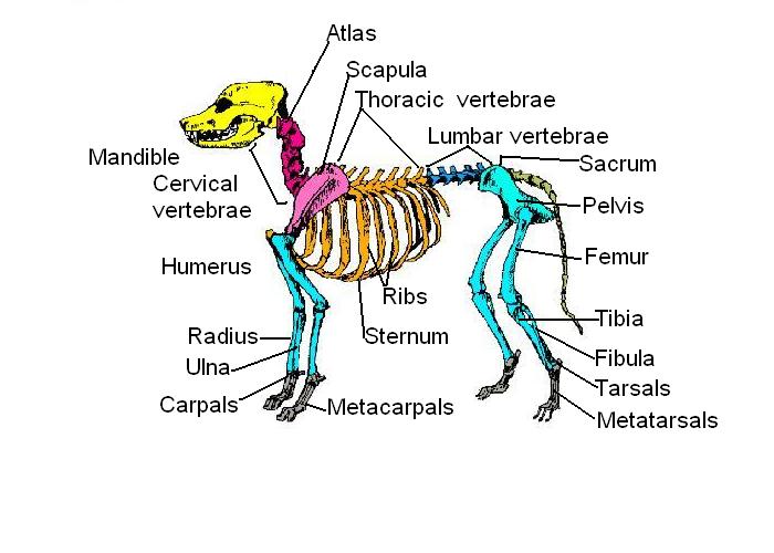
Dog Skeletal Anatomy - Exploring Nature Animal Anatomy (Veterinary Diagrams) Dog Skeletal Anatomy. High Resolution PDF for Printing. Click Here. Link to More Information About This Animal. Click Here. Citing Research References. When you research information you must cite the reference. Citing for websites is different from citing from books, magazines and periodicals. How to Work Safely with - Hazardous Products using the "Skull and ... The symbol within the pictogram is a human skull with two crossed bones behind it. The symbol indicates that hazardous products with this pictogram can cause death or poisoning. Hazardous products with this pictogram can be safely worked with if proper storage and handling practices are followed. Diagram of Human Heart and Blood Circulation in It A heart diagram labeled will provide plenty of information about the structure of your heart, including the wall of your heart. The wall of the heart has three different layers, such as the Myocardium, the Epicardium, and the Endocardium. Here's more about these three layers. Epicardium Estrogen - Wikipedia Estrogen or oestrogen is a category of sex hormone responsible for the development and regulation of the female reproductive system and secondary sex characteristics. There are three major endogenous estrogens that have estrogenic hormonal activity: estrone (E1), estradiol (E2), and estriol (E3). Estradiol, an estrane, is the most potent and prevalent. ...
Anatomy - LibGuides at Duke University Medical Center Visible Body - Skeleton. Muscolosketal MRI. Hypermuscle: Muscles In Action. Master Muscle List. Muscle Atlas. HeadNeckBrainSpine. Autonomic Nervous System Tutorial. Interactive Neuroanatomy Atlas. NeuroAnatomy Web Resources. The Whole Brain Atlas. Atlas of Human Anatomy by Frank H. Netter. ISBN: 9780808924517. Stegosaurus - Wikipedia Stegosaurus (/ ˌ s t ɛ ɡ ə ˈ s ɔːr ə s /; lit. 'roof-lizard') is a genus of herbivorous, four-legged, armored dinosaur from the Late Jurassic, characterized by the distinctive kite-shaped upright plates along their backs and spikes on their tails. Fossils of the genus have been found in the western United States and in Portugal, where they are found in Kimmeridgian- to early Tithonian ... skeleton coloring pages anatomy - Just As Much Fun Log-Book Diaporama Enhance your Skeletal System Unit with these Skeleton Doodle Notes. Here is the list of printable skeleton coloring pages. This worksheet includes a coloring page for students to create a key and color in the skeleton so they can individually identify what category each bone falls into. Science from Scratch - Anatomy and Biology. World of Biology. Kidney Structures and Functions Explained (with Picture and Video) The glomerulus connects to a long, convoluted renal tubule which is divided into three functional parts. These consist of the loop of Henle (nephritic loop), the proximal convoluted tubule, and the distal convoluted tubule, which empties into the collecting ducts. These collecting ducts fuse together and enter the papillae of the renal medulla.
Diagrams - Skeletal System
List of skeletal muscles of the human body - Wikipedia The terms "artery" and "nerve" are both used when these structures are mentioned. Contents 1 Head 1.1 Forehead/eyelid 1.2 Extraocular muscles 1.3 Ear 1.4 Nose 1.5 Mouth 1.6 Mastication 1.7 Tongue 1.7.1 Extrinsic muscle 1.7.2 Intrinsic 1.8 Soft palate 1.9 Pharynx 1.10 Larynx 2 Neck 2.1 Clavicular 2.2 Suprahyoid 2.3 Infrahyoid 2.4 Neck 2.4.1 Anterior

frontal sinus | Anatomy System - Human Body Anatomy diagram and chart images
Antenatal Care Module: 6. Anatomy of the Female Pelvis and Fetal Skull 6.1.1 Ilium. Ilium is pronounced 'ill ee umm' and iliac is 'ill ee ack'. The major portion of the pelvis is composed of two bones, each called the ilium — one on either side of the backbone (or spinal column) and curving towards the front of the body. When you place your hand on either hip, your hand rests on the iliac crest, which is the upper border of the ilium on that side.
Skeletal System Diagram Labeled For Kids - Diagram Media
Creating Model Working Lungs: Just Breathe - Activity Peel off the labels, if any, on the 2-liter bottles. Tell students that the 2-liter bottle represents the human chest cavity. Stick two drinking straws through the two holes in the bottle cap. Place one 9-inch balloon on the end of each straw and secure them with rubber bands, as shown in Figure 2. Figure 2.
Human skeletal system - Teaching resources
Centriole - National Human Genome Research Institute Home A centriole is a barrel-shaped organelle which lives normally within the centrosome. The centrosome is the area of the cytoplasm. It's next to the nucleus and within the centrosome. The word some refers generally to an organelle of some sort, like a lysosome or an endosome. Within that centrosome there are two centrioles.
Anatomy & Physiology - Skeletal System
Anatomy Of The Lower Extremity Veins - Varicose Veins The posterior accessory GSV of the leg (Leonardo's vein or posterior arch vein) is a common tributary, it begins posterior to the medial malleolus, ascends on the posteromedial aspect of the calf, and joins the GSV distal to the knee (see Figure 2.8). The anterior accessory GSV of the leg drains the anterior aspect of the leg below the knee.

Skeletal System Diagram 8-Pack by Not Weird Homeschoolers | TpT
Step 1 Organize bones three different ways. Think about the ... Step 1 Organize bones three different ways. Think about the organization of the bones in the human body and outline three different ways in which the skeletal system is organized. Use a visual diagram or chart to present your three organizational schemes. Be detailed and very clear in the presentation of your ideas.

Skeleton With Scientific Labels - Home schooling with A2Z Home's Cool | A2Z Homeschooling
Spinal Cord Cross Section Explained (with Videos ... - New Health Advisor Looking at a cross section of the spinal cord, you would see gray matter shaped like a butterfly surrounded by white matter. The gray matter is the core and ends up to be four projections that are known as horns. At the back are two dorsal horns and away from the back are two ventral horns.

Skeletal System Diagrams for Labeling, With Reference Information and Charts
Bivalvia - Wikipedia Bivalvia (/ b aɪ ˈ v æ l v i ə /), in previous centuries referred to as the Lamellibranchiata and Pelecypoda, is a class of marine and freshwater molluscs that have laterally compressed bodies enclosed by a shell consisting of two hinged parts. Bivalves as a group have no head and they lack some usual molluscan organs like the radula and the odontophore.They include the clams, oysters ...
Skeletal System Diagram 8-Pack by Not Weird Homeschoolers | TpT
Organs of Skeletal System and Their Functions - New Health Advisor Vertebral column - consist of all spinal vertebrae. Thoracic cage - it contains ribs and sternum. Appendicular Skeleton It contains the following from top to bottom respectively: Shoulder girdle - it includes clavicle and scapula. Upper limb - it contains arm, forearm and hand's bones. Hip girdle - it includes hip bone.
Medical infographic : Human Skeleton | Humans | Now Science News - InfographicNow.com | Your ...
The Biomechanics of Human Skeletal Muscle - MHMedical.com This characteristic enables skeletal, or striated, muscle to perform the important functions of maintaining upright body posture, moving the body limbs, and absorbing shock. Because muscle can only perform these functions when appropriately stimulated, the human nervous system and the muscular system are often referred to collectively as the ...

Skeletal System Front : Biological Science Picture Directory – Pulpbits.net
Functions of Human Skeletal System | Just-Health.net It is part of the skeletal system, which is composed of ligaments and cartilages, in addition to bones. Bones are connective tissues made of osseous materials that undergo calcification, a process where minerals are deposited to harden the bone. Cartilages are thick and rubbery tissues that are found in joints, the ears, the nose, and the ribs.

THE MUSCULOSKELETAL SYSTEM worksheet - Free ESL printable worksheets made by teachers
Sequence Diagram Tutorial - Complete Guide with Examples A sequence fragment is represented as a box that frames a section of interactions between objects (as shown in the examples below) in a sequence diagram. It is used to show complex interactions such as alternative flows and loops in a more structured way. On the top left corner of the fragment sits an operator.

Dog Anatomy: Canine 3D for Windows 8 and 8.1
Cardiovascular System Worksheet Pdf - flakeinspire Cardiovascular system diagrams, quizzes and free worksheets. 1 cardiovascular system components of the cardiovascular system • consists of the heart plus all the blood vessels • transports blood to all parts of the body in two 'circulations': P 14 of a study guide for 4th grade via westerndairyassociation org wp wp 06 skeletal system pdf ...

muscular pictures 744×1179 | Anatomy System - Human Body Anatomy diagram and chart images
The Anatomy and Physiology of Animals/Skeleton Worksheet/Worksheet Answers - WikiEducator
Post a Comment for "44 skeletal system diagram with labels"