39 fluorescent labels and light microscopy
Light Sheet Fluorescence Microscopy - an overview ... Applications of single-molecule fluorescence microscopy. (A) The photophysical properties of a fluorophore contain information about its position and its state. This allows, for example, tracking molecules, observing conformational and constitutional changes, or following chemical reactions. (B) Examples for applications in biology and chemistry. In Silico Labeling: Predicting Fluorescent Labels ... - Cell Fluorescence microscopy images can be predicted from transmitted-light z stacks • 7 fluorescent labels were validated across three labs, modalities, and cell types • New labels can be predicted using minimal additional training data Summary Microscopy is a central method in life sciences.
Light microscopy of proteins in their ultrastructural context by O M’Saad · 2020 · Cited by 34 — Fluorescence microscopy has transformed the field of cell biology through its exceptional contrast and high specificity of labeling.
Fluorescent labels and light microscopy
Visualizing the invisible: New fluorescent DNA label ... A molecule of interest is labelled with a special fluorescent dye that flashes on and off like a blinking star. Unlike traditional fluorescence microscopy, which uses labels that glow constantly,... Fluorescence Microscopy Sir George Stokes, a British scientist, first discovered fluorescence in 1852 when he observed that the mineral fluorite (Fig. 1, molecular formula CaF2) emitted red light when it was illuminated by ultraviolet excitation. Early investigations in the 19th century showed that many specimens (including minerals, crystals, drugs, butter, chlorophyll, and vitamins) fluoresce when irradiated with… Label-free imaging of live cells - CytoSMART Study live cells using non-invasive & non-toxic methods Live-cell imaging has become a fundamental tool to better understand cellular processes and biological functions. A non-invasive and non-toxic alternative to fluorescent microscopy is label-free imaging. Although the resolution achievable…
Fluorescent labels and light microscopy. Fluorescence Microscopy vs. Light Microscopy Light microscopy does much what the name implies: visible light and magnifying lenses are used to view small objects. Light microscopes are the oldest form of higher quality imaging devices, dating back to the 1500s, and were the microscopes with which first cells were observed. A common method to visualize cells or tissue with light microscopes is... Label-free prediction of three-dimensional fluorescence ... Fluorescence microscopy can resolve subcellular structure in living cells, but is expensive, slow, and toxic. Here, we present a label-free method for predicting 3D fluorescence directly from transmitted light images and demonstrate its use to generate multi-structure, integrated images. Fluorescent tag - Wikipedia Fluorescent tag. S. cerevisiae septins revealed with fluorescent microscopy utilizing fluorescent labeling. In molecular biology and biotechnology, a fluorescent tag, also known as a fluorescent label or fluorescent probe, is a molecule that is attached chemically to aid in the detection of a biomolecule such as a protein, antibody, or amino acid. Illuminating Life: Fluorescence Microscopy - The Scientist Illuminating Life: Fluorescence Microscopy. A 175-year journey of discovery fashioned fluorescence microscopy into a fundamental technique for life science research. Fluorescent and phosphorescent labels in the form of protein tags and dyes enable researchers to visualize almost any cellular process.
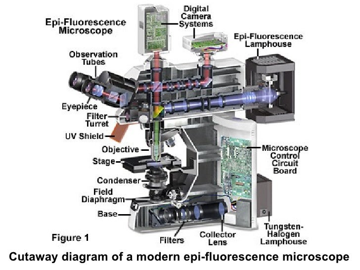
Fluorescence Microscopy - an overview | ScienceDirect Topics In most modern fluorescence microscopes, epi-illumination is employed. This means that the light used for excitation is reflected onto the specimen through the ... Labeling the ER for Light and Fluorescence Microscopy by C Hawes · 2018 · Cited by 3 — The ER is a highly dynamic network of tubules and membrane sheets. Hence imaging this organelle in its native and mobile state is of great importance. Light Microscope- Definition, Principle, Types, Parts ... A minimum distance (d) between two objects that distinguishes them to be two separate entities, determined by the wavelengths of the light can be calculated by an Abbe equation using the wavelength of the light that illuminated the specimen (Lambda, λ) and the numerical aperture (NA, n sin Ɵ) i.e. d=0.5 λ/n sin Ɵ A quick guide to light microscopy in cell biology - PMC - NCBI Fluorescence microscopy uses fluorescent dyes (fluorophores), which are molecules that absorb one wavelength of light (the excitation wavelength) and emit a second, longer wavelength of light (the emission wavelength).
Evanescent Light-Scattering Microscopy for Label-Free ... Measurements and theoretical analysis revealed that light-scattering signals originating from single surface-bound lipid vesicles enable characterization of their sizes without employing fluorescent lipids as labels. Fluorescence Microscopy - Explanation and Labelled Images Dec 16, 2020 · Fluorescence microscopy uses a high-intensity light source that excites a fluorescent molecule called a fluorophore in the sample observed. The samples are labeled with fluorophore where they absorb the high-intensity light from the source and emit a lower energy light of longer wavelength. Fluorescence Microscope: Principle, Types, Applications Fluorescence microscopy is a type of light microscope that works on the principle of fluorescence. A substance is said to be fluorescent when it absorbs the energy of invisible shorter wavelength radiation (such as UV light) and emits longer wavelength radiation of visible light (such as green or red light). Different Ways to Add Fluorescent Labels | Thermo Fisher ... Using fluorescence provides greater contrast compared to viewing your samples with brightfield microscopy alone. Labeling various targets with separate fluorescent colors allows you to visualize different structures or proteins within a cell in the same experiment.
FLUORESCENCE MICROSCOPY: A program to help labs 'go green' | Laser Focus World
Fluorescence Microscopy & Cell Imaging - UNM Health The Fluorescence Microscopy and Cell Imaging Shared Resource provides UNM researchers access to state-of-the art instrumentation for multiple fluorescence and transmitted light microscopy techniques: Laser scanning single and multi-photon microscopes and hyperspectral imaging systems enable simultaneous visualization and quantification of ...
Fluorescence Microscopy
Fluorescent Labelling - ScienceDirect.com Fluorescence microscopy Fluorescent labeling methods are generally based on reactive derivatives of fluorophores that selectively bind to functional groups contained in target biomolecules and are widely used in biotechnology because of their non-destructive properties and the high sensitivity of fluorescence techniques ( Sahoo, 2012 ).

A schematic diagram of the light path in the ptychographic microscope... | Download Scientific ...
Imaging Flies by Fluorescence Microscopy: Principles ... The development of fluorescent labels and powerful imaging technologies in the last two decades has revolutionized the field of fluorescence microscopy, which is now widely used in diverse scientific fields from biology to biomedical and materials science. ... has brought about the era of fluorescence light microscopy. The first fluorescence ...

טוטו יחס קבוע - JHaringan
Fluorescent labeling of abundant reactive entities (FLARE ... Fluorescence microscopy is a technique that is commonly used in the biomedical sciences. It offers the powerful ability to visualize structures or molecules in three dimensions within biological...

Label-free fluorescence microscopy offers early cancer detection – Physics World
A quick guide to light microscopy in cell biology FLUORESCENCE MICROSCOPY · by K Thorn · 2016 · Cited by 134 — Most molecules in the cell are not very fluorescent, so fluorescent labels to be imaged are typically introduced by the ...

Light sheet fluorescence microscopy for Multiview imaging of living and cleared specimens ...
How do you fluorescently label mRNA for microscopy? However, you can also do fluorescence in situ hybridization (FISH) to label your mRNA inside of the cells, but this is a much more laborious process. Cite 11th Jun, 2021

FBI Workshop: Light-sheet Fluorescence Microscopy – Principles and Systems Overview – France ...
Light sheet fluorescence microscopy | Nature Reviews ... Light sheet fluorescence microscopy (LSFM) is a technique that uses a thin sheet of light for illumination, allowing optical sectioning of the sample. In this Primer, Stelzer et al. outline the ...
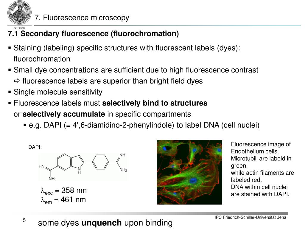
PPT - 7.1 Fluorochromes Fluorescence microscopy differentiates between two kinds of ...
Rethinking Fluorescence Microscopy | Lab Manager One such piece of laboratory equipment is the fluorescence microscope. Although a familiar sight in many life science laboratories, the light source can easily be overlooked. Many microscopes still use energy-hungry, toxic mercury or metal halide lamps, but modern LED technology now offers a cleaner, more efficient, and high-performance ...

Label-free prediction of three-dimensional fluorescence images from transmitted-light microscopy ...
Novel Fluorescent Label Shines a Light on DNA Structure in ... Liu and her team formulated a new label called Hoechst-Cy5 to overcome the hurdle that fluorescent dyes didn't work well on DNA or in processed clinical cancer samples. After showing that the new...

Design of a dynamic biofilm imaging cell for white-light interferometric microscopy
New fluorescent label provides a clearer picture of how ... A molecule of interest is labelled with a special fluorescent dye that flashes on and off like a blinking star. Unlike traditional fluorescence microscopy, which uses labels that glow constantly,...
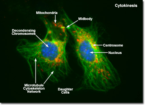
Molecular Expressions Cell Biology: Mitosis with Fluorescence Microscopy - Cytokinesis
Fluorescent Label - an overview | ScienceDirect Topics 14.3.2Fluorescence quenching microscopy Fluorescence microscopy is a very common tool. Usually, fluorescent labelsare used to brighten up the object of interest. However, the same strategy is not applicable for graphitic materials, such as graphite, graphene, GO or r-GO as they are strong quenchers of dye molecules [45].
Cleared Sample Imaging by Means of Light-Sheet Fluorescence Microscopy | Bruker
Light Microscopy in Trypanosomes: Use of Fluorescent ... Spray a glass microscope slide with 70% (v/v) ethanol and wipe clean. Pipet 3 μL of cell solution onto the glass slide and carefully place a No. 0 coverslip (50 × 22 mm) on top of the cell solution. 6. Check the slide on an inverted microscope using a 20× objective to ensure that the cells are well immobilized on the slide ( see Note 7 ). 7.
Researchers design wearable microscope that can measure fluorescent dyes through skin
Introduction to Fluorescence Microscopy | Nikon's MicroscopyU In fact, the fluorescence microscope is capable of revealing the presence of a single molecule. Through the use of multiple fluorescence labeling, ...

Nature Methods: Light-sheet Fluorescence Microscopy - Method of the Year 2014 | Learn & Share ...
Fluorescence Imaging - Teledyne Photometrics Fluorescent molecules (known as fluorophores) are used to label samples, and fluorophores are available that emit light in virtually any color. In a fluorescent microscope, a sample is labeled with a fluorophore, and then a bright light ( excitation light) is used to illuminate the sample, which gives off fluorescence ( emission light ).

Fluorescence Imaging
Fluorescent Microscopy - SERC - Carleton Jan 15, 2021 — A fluorescence microscope is much the same as a conventional light microscope with added features to enhance its capabilities.

Label-free prediction of three-dimensional fluorescence images from transmitted-light microscopy ...
Label-free prediction of three-dimensional fluorescence ... Label-free prediction of three-dimensional fluorescence images from transmitted-light microscopy Understanding cells as integrated systems is central to modern biology. Although fluorescence microscopy can resolve subcellular structure in living cells, it is expensive, is slow, and can damage cells.
Post a Comment for "39 fluorescent labels and light microscopy"